To begin with, it is necessary to define what histology is. Translated from ancient Greek, it sounds like "the science of tissues." But this name is not entirely accurate and narrows the scope of its activities, since the methods of histology study not only living tissues, but also the fine structure of organs, and even cells.
In addition, histology is a science that studies the evolution of tissues and cells, their development and formation in the body, the work of tissues, cells and organs, and intercellular substance. Histology also investigates the regeneration of tissues, which provides them with functional and structural integrity.
History of histology
Histology appeared much earlier than the microscope. Fabrics were described by Aristotle, Avicenna, Galen, Vesalius. However, the concept of a cell was introduced only by R. Hooke in 1665 after he examined the cellular structure of plant tissue under a microscope. A number of scientists carried out the first histological studies, as a result of which, thanks to the efforts of K. Wolf and K. Baer, a new branch appeared – embryology.

By the nineteenth century, histology had become a real academic science. In the middle of the century, the modern theory of tissues was laid, the science of tissue and cellular pathology began to develop. The development of histology was pushed by the creation of the cell theory and the next discoveries in cytology. A great contribution to the development of this science was made by such luminaries as I. Mechnikov and L. Pasteur, who laid the foundation for the doctrine of the immune system.
There were also curiosities: the histologists K. Golgi and S. Ramon y Cajal interpreted the same images of the brain section in different ways and came to opposite assumptions regarding its structure, which did not prevent both from receiving the Nobel Prize in Medicine in 1906 .
The methodology of histology continued to improve in the last century, as a result of which this science has acquired its current shape. Now it is closely intertwined with cytology, medicine, embryology and other disciplines. It deals with such issues as adaptation at the tissue and cellular level, differentiation of tissues and cells and the patterns of their development, regeneration of organs and tissues, etc. Achievements in pathohistology are widely used in medical practice, as they facilitate understanding of the mechanism of the disease and the search for effective therapy.
Sections of histology
There are three sections in this discipline:
- general histology;
- cytology ;
- private histology or microscopic anatomy.
It is known that cytology is a science that studies cells – the elementary bricks that make up and thanks to which all living matter on Earth functions. The task of general histology is to study the origin, structure, functioning and development of tissues. There is also a private histology, which studies the structure of organs at the microscopic and ultramicroscopic levels. Some artificiality of dividing histology into these sections should be recognized, since in reality tissues are formed from cells that make up organs, and the totality of the latter forms an organism. Therefore, parts of the body are both organs and tissues with cells. But still, such a division of histology is justified by the fact that it is easier to present the material, talking about what histology studies.
Video about what histology studies
Since histology is the science of living objects, its development is unthinkable without close contact with other biological sciences: anatomy, genetics, physiology, embryology, and others. It also needs a connection with physics and chemistry, since in its research histology constantly uses numerous chemical reagents (dyes, fixatives), physicochemical research methods, and physical instruments (microtomes, microscopes).
Histological studies
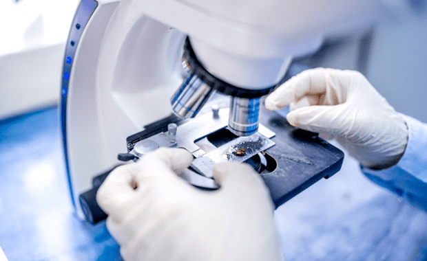
The main histological method of research remains microscopic. Preparations first undergo a certain preparation, after which they are examined under a microscope. When preparing the preparation, the thinnest sections are made from it, which are then stained with a suitable dye and fixed. And then the preparations are studied under a microscope.
Histological studies can also be carried out on living preparations, although the study of a living object is rather difficult, since in transmitted light the histological structures are colorless and poorly distinguishable in the microscope field, and besides, due to their large size, they simply cannot be placed under a microscope. In this regard, the study of fixed objects prevails, that is, already dead, specially processed cells, but retaining their chemical composition and structure. Each method has advantages and disadvantages, so they try to use them together so that they complement each other.
What histology studies in biology among living structures is greatly assisted by modern technology. For example, the chemical composition and physical properties of cells are studied on living objects, on which a number of operations can be performed using a micromanipulator, such as transplanting nuclei from cell to cell, removing intracellular structures, etc.

Histological research methods
The main methods of histological studies include:
- Optical microscopy, which studies miniature histological specimens using a variety of optical microscopes, including those with radiation sources with different wavelengths. It is known that in a conventional microscope the light source is sunlight or artificial light with a minimum wavelength of 0.4 μm.
- Dark-field microscopy is based on the fact that only the radiation obtained by diffraction on the preparation structure reaches the objective. To do this, a condenser is built into the microscope, which sends a strictly oblique beam of light from the side to the preparation. In this case, the field of the microscope remains dark, and only small particles of the preparation reflect an oblique beam that reaches the objective.
- Phase contrast microscopy.
- Fluorescent and luminescent microscopy. There are a number of substances whose molecules or atoms are capable of absorbing short-wave radiation, passing into an excited state. During the reverse transition from the excited to the normal state, the atom again emits a photon, but with a longer wavelength.
- interference microscopy. In such a microscope, the beam of light received from the lighting lamp is divided into two streams: the first beam passes through the object, changing the phase of the oscillations, and the second goes directly to the lens without changing the phase. Both beams are then combined in the prisms of the objective, resulting in their interference. In the lens, an image is obtained in which sections of the sample under consideration, which have different optical density and thickness, acquire different contrast. Once quantified, the mass and dry matter concentration can be calculated.

-
Electron microscopy has become a revolutionary step in the development of microscopy. Both TEM transmission electron microscopes, which transmit light through a sample, and SEM scanning or scanning electron microscopes, operating on the scattering effect, were created and are now actively used for research. In transmission microscopes, only a two-dimensional image of the microobject under study can be obtained, and in order to obtain a spatial representation of the structure under study, SEM is used, which gives a three-dimensional picture. A scanning electron microscope acts as an electron microprobe that scans the object under study: it “feels" all points on the surface with a narrowly focused electron beam in succession. To survey the selected area, the microprobe moves over the surface of the sample due to the action of deflecting coils (similar to a television sweep). Therefore, the study is called scanning or reading, and the field along which the microprobe moves is called a raster. The resulting signal is displayed on the monitor screen, the movement of the electron beam of which is synchronized with the beam of the microprobe.
-
Ultraviolet microscopy uses lamps that emit ultraviolet radiation having a wavelength of 0.2 microns.
-
polarizing microscopy.
-
Radio autography.
-
Cytospectrophotometry.
-
Cell culture method.
-
immunocytochemical methods.
-
Cell microsurgery.