What is microtomography?
Currently, there are not many methods for non-invasive reconstruction of three-dimensional models representing the internal structure of the object under study. Developments are underway in this area, this type of research is of particular interest to the scientific community in different countries, as it can provide a competitive advantage for certain industries. One of the above methods is computed microtomography (you can often find such a name as x-ray microtomography, these are two terms denoting the same analysis method, the first name often appears in foreign literature). Let us consider this type of analysis of the internal environment in more detail.
The history of the discovery of microtomography
Research in the field of this method has been carried out for quite a long time, in Russia the first use of microtomography is attributed to the beginning of the nineties of the last century, and it was in Russia that the first study was carried out on the analysis of minerals, ores and rocks, which immediately served as an impetus for the formation of the applied nature of microtomographic analysis in our country. The first studies were carried out on the basis of such scientific institutions as Moscow State University named after M.V. Lomonosov and the Russian Academy of Sciences, which made it possible to immediately involve a huge number of specialists and inform a wide variety of industries about the new direction.
Already from the very inception of the method, its possible application for a wide range of various studies has become obvious. Initially, active research was carried out in the oil and gas sector, but then the undeniable advantages of the method were appreciated in such areas as:
- biology;
- medicine;
- chemistry;
- archeology;
- soil science;
- microelectronics.
At the moment, research in the field of studying the volumetric structure of objects using X-ray microtomographs continues to develop, more and more domestic enterprises are acquiring similar equipment for themselves, in 2012 there were 40 research teams in the country, at the moment their number has almost doubled.
Such a rapid development and distribution becomes possible due to the fact that X-ray microtomography is considered a very promising direction, since the results obtained are really necessary and can significantly advance a number of experiments.
Watch a video about the creation of a domestic microtomograph by Russian scientists.
How does a microtomograph work?
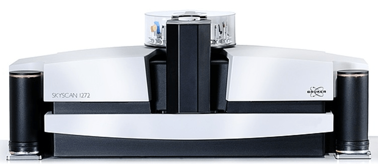
The construction of equipment for this type of analysis is very complex and requires not only the high quality of the materials from which it is made, but also highly qualified specialists who could design a truly reliable device. Only a few companies in the world are developing this type of equipment, the most popular of which is Bruker microCT, formerly this company was called Skyscan. It was founded in Belgium by a Russian scientist from Lomonosov Moscow State University, and now this company is widely known throughout the world, produces many types of tomographs, and also supplies special software necessary to work with them.
The basis of a microtomograph is a micro- or in some cases even a nano-sized tube that shines through the object under study with X-rays, and a special camera installed nearby that receives X-rays receives shadow projections of an enlarged size. The object inside rotates on delivery, at this time the X-ray camera receives and records several hundred shadow projections, which are subsequently processed by a computer. Based on the obtained projections, the computer builds a set of virtual models representing sections of the object.
After processing the results, it is possible to view all kinds of sections of the object, they can be flipped and rotated, as well as to obtain all the necessary measurements and numerical characteristics of the entire volume of the internal environment of the object or even its separate area. This in combination allows you to create three-dimensional models that provide opportunities for virtual movement through the internal environment of the object of study.
X-ray microtomography: advantages
With the discovery of microtomography, the understanding of the internal structure of many types of rocks, oil reservoirs, and various composite polymers has radically changed; these studies have been the foundation for further research, improvements in geology, physics, and chemistry.
The main advantages of this research method are:
It does not require any preliminary preparation, which significantly reduces the time for analysis and obtaining the result. There is no need to pre-prepare samples, pre-stain specimens, or obtain thin sections.


